What happens when an enzyme of cortisol synthesis is missing?
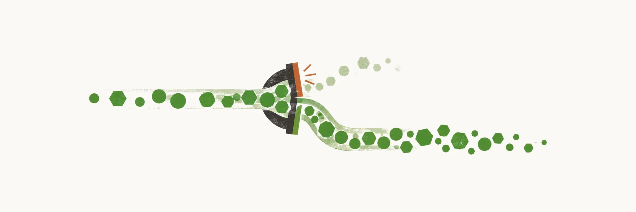
Cortisol is built step by step in the cells of the zona fasciculata, like a production line, and five key enzymes carry out the steps. A mutation in one of these enzymes blocks the line at that step. Everything before the block piles up and everything after it falls. The pile-up then spills into any side branch of the line that is still open.
The most common mutation affects 21-hydroxylase (P450 c21). Its substrate in the glucocorticoid pathway is 17-hydroxyprogesterone (17-OHP), so when 21-hydroxylase activity is missing or reduced, 17-OHP accumulates in front of the block.
Cortisol falls as well. As the overview of this condition describes, a block in cortisol synthesis removes negative feedback on ACTH, so ACTH becomes very high in these patients and drives hyperplasia of the adrenal cortex.
Why does the pile-up cause virilization?
The accumulated 17-OHP has nowhere to go but the androgen pathway, which is still intact, and the high ACTH keeps driving the cortex. The accumulating precursors are diverted into androgens, so the accumulated 17-OHP is shunted into the androgenic pathway. Androgen over-production causes virilization, the development of male characteristics. That is why a child with this block is short of cortisol but has too many androgens.
High ACTH has a second effect: it causes hyperpigmentation. ACTH and beta-lipotropin both have melanocyte-stimulating activity.
Both effects come from the same block, but not every patient has it to the same degree. Why do patients differ?
Why do different patients have different severity?
The severity of the block determines the form of the disease. Different mutations leave different amounts of residual 21-hydroxylase activity, and the level of enzymatic activity is correlated with the onset of the disease.
21-hydroxylase deficiency (21OHD) CAH therefore comes in two forms, classical and non-classical. The classical form was described first. It has an early onset, at birth, which fits a severe block. Each person carries two copies of the gene, and the classical form tends to result from two severely affected genes.
The non-classical form, also known as late-onset CAH, was described later. Its onset is later, during adolescence, because its mutations leave a low level of 21-hydroxylase activity. It tends to result from two mildly affected genes, and it may also result from one severely affected and one mildly affected gene. The non-classical form is more common than the classical form.
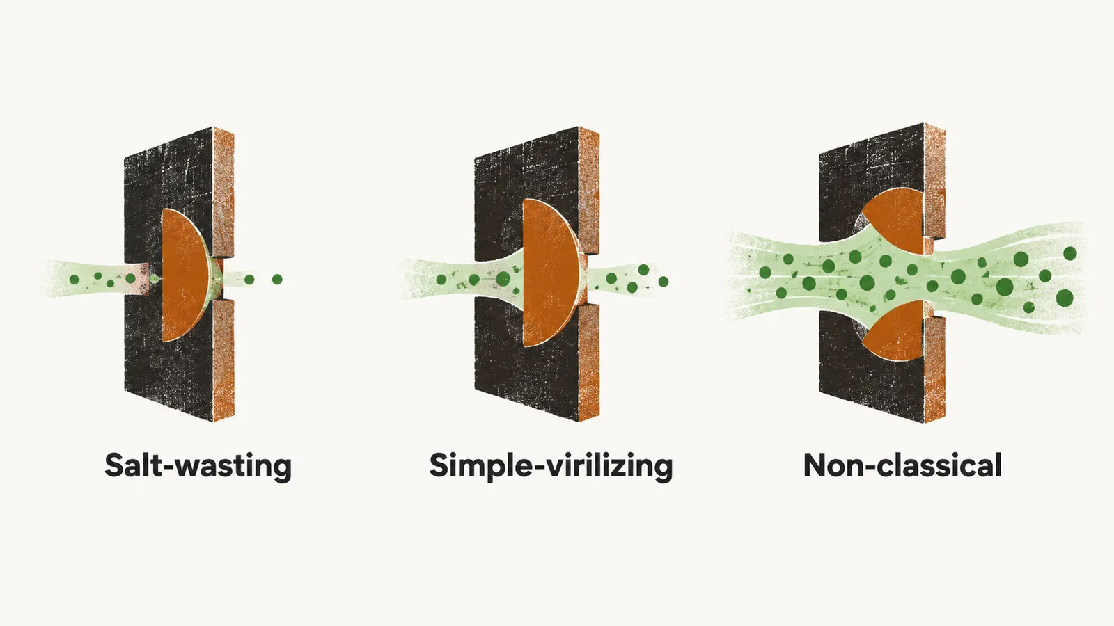
Within the classical form, the amount of enzyme left separates two types. The salt-wasting type leaves almost no 21-hydroxylase activity, whereas the simple-virilizing type leaves only a little. What the difference costs is explained by the aldosterone route.
Where does salt wasting come from?
21-hydroxylase also sits on the route that makes aldosterone. A severe lack of 21-hydroxylase, as seen in classical 21OHD CAH, can therefore interfere with the normal mineralocorticoid production pathway. The result of aldosterone deficiency in these patients is called salt wasting. Aldosterone is needed for the distal tubules to reabsorb sodium, so without enough of it the kidney loses sodium.
Salt wasting happens mainly because of the lack of aldosterone. Another mechanism can exacerbate it: an antagonistic effect of accumulated 17-OHP on the aldosterone receptors in the distal convoluted tubules.
A little residual enzyme protects aldosterone, because in vitro studies show that only 1% activity is enough for the formation of enough aldosterone. Salt wasters are the patients with a complete lack of 21-hydroxylase activity, the severely affected CAH phenotypes with almost no residual activity.
Severity also matters before birth, in the fetus. How does it affect a developing child?
Why are females, but not their internal genitalia, affected before birth?
Adrenocortical activity begins in fetal life, at the 7th week of gestation. The critical time for sexual differentiation is the 9th to 12th week of gestation, so the adrenal is already making hormones when the external genitalia are being shaped. Female patients with 21-hydroxylase deficiency accumulate androgen, and because of this non-physiological state their external genitalia become virilized. The development of the female external genitalia can be affected by the androgen level.
The internal genitalia (uterus, fallopian tubes and ovaries) are not affected by androgen. Their development is Mullerian-dependent. In CAH patients no anti-Mullerian hormone (AMH) is present, and AMH is produced by Sertoli cells. So the Mullerian structures are not removed and keep their normal form.
Male patients show small testes. Their gonadotropin level is low, suppressed by the high androgen level.
What do blocks at other steps look like?
Other enzymes along the same pathway can also be defective, and each gives a distinct picture. The same logic applies. A block at one of the earliest steps stops every steroid, because StAR and the side-chain cleavage enzyme P450scc lie at the start of the chain, so all products are low. A block later in the chain loses fewer hormones but lets precursors with their own actions pile up; for example, deoxycorticosterone (DOC) acts on the mineralocorticoid receptor, which explains the rows with hypokalemic low-renin hypertension. P450 c17 carries out both 17α-hydroxylation and 17,20-lyase activity, so its loss affects cortisol and sex steroids at once. P450 oxido-reductase supplies several P450 enzymes (21-hydroxylase, 17-hydroxylase and aromatase), which is why its deficiency causes partial, combined and variable defects. The 21-hydroxylase rows repeat the picture above.
| Defect (enzyme or protein) | Onset | Genitalia | Mineralocorticoid effect | Typical features | Gene |
|---|---|---|---|---|---|
| Lipoid CAH, StAR protein | Congenital | Female genitalia with no sexual development | Salt wasting | All steroid products low | StAR, 8p11.2 |
| Lipoid CAH, P450scc | Congenital | Female genitalia with no sexual development | Salt wasting | All steroid products low | CYP11A, 15q23-24 |
| 3β-HSD deficiency, classic | Congenital | Females virilized, males under-virilized | Salt wasting | Elevated DHEA, 17-pregnenolone; low androstenedione, testosterone; elevated K, low Na, CO2 | HSD3B2, 1p13.1 |
| 3β-HSD deficiency, non-classic | Postnatal | Normal genitalia, with mild to moderate hyperandrogenism postnatally | None | Elevated DHEA, 17-pregnenolone; low androstenedione, testosterone | Absent or unknown |
| 17α-OH deficiency (P450c17) | Congenital | Variable sexual development | Hypokalemic low-renin hypertension | Normal or decreased androgens and estrogen; elevated DOC, corticosterone | CYP17, 10q24.3 |
| 17,20-Lyase deficiency (P450c17) | Congenital | Infantile female genitalia | None | Decreased androgens and estrogens | CYP17, 10q24.3 |
| Combined 17α-OH/17,20-lyase deficiency, congenital (P450c17) | Congenital | Infantile female genitalia | Hypokalemic low-renin hypertension | Decreased androgens and estrogens | CYP17, 10q24.3 |
| Combined 17α-OH/17,20-lyase deficiency, postnatal (P450c17) | Postnatal | Infertility, infantile female genitalia | None | Decreased follicular estradiol, increased progesterone | CYP17, 10q24.3 |
| Classic 21-OH deficiency, salt wasting (P450c21) | Congenital | Females prenatally virilized, normal male genitalia; hyperpigmentation | Salt wasting | Elevated 17-OHP, DHEA, androstenedione; elevated K, low Na, CO2 | CYP21, 6p21.3 |
| Classic 21-OH deficiency, simple virilizing (P450c21) | Congenital | Females prenatally virilized, normal male genitalia | None | Elevated 17-OHP, DHEA, androstenedione; normal electrolytes | CYP21, 6p21.3 |
| Non-classic 21-OH deficiency (P450c21) | Postnatal | Males and females with normal genitalia at birth, hyperandrogenism postnatally | None | Elevated 17-OHP, DHEA, androstenedione on ACTH stimulation | CYP21, 6p21.3 |
| Classic CAH, 11β-deficiency (P450c11B1) | Congenital | Females virilized with atypical genitalia, males unchanged | Low-renin hypertension | Elevated DOC, 11-deoxycortisol (S), androgens; low K, elevated Na, CO2 | CYP11B1, 8q24.3 |
| Non-classic CAH, 11β-deficiency (P450c11B1) | Postnatal | Males and females with normal genitalia at birth, hyperandrogenism postnatally | None | Elevated 11-deoxycortisol ± DOC, elevated androgens | CYP11B1, 8q24.3 |
| P450 oxido-reductase deficiency (POR) | Congenital | Females virilized with atypical genitalia, males under-virilized | None | Partial, combined and variable defects of P450c21, P450c17 and P450aro activity | POR, 7q11.2 |
All of these changes first show in a child. How do they appear in the genitalia, growth, puberty and electrolytes?