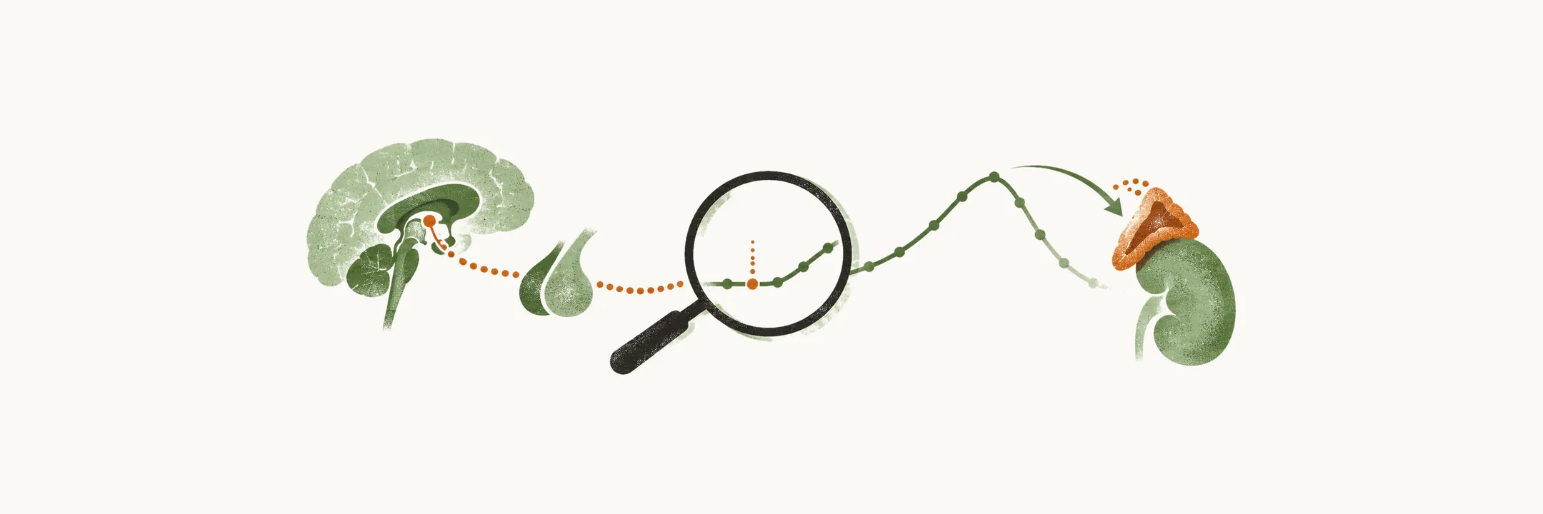
The adrenal cortex is the last link in a chain: CRH from the hypothalamus drives ACTH from the pituitary, and ACTH drives cortisol from the cortex. Diagnosing adrenal insufficiency means answering three questions in turn. Is cortisol really too low for the situation? Which link in the chain has failed? What damaged that link?
What does the diagnosis of adrenal insufficiency have to establish?
The diagnosis rests on laboratory confirmation of inappropriately low cortisol secretion. Then the insufficiency is classified as primary, secondary or tertiary. Symptoms are non-specific, so they cannot make the diagnosis alone.
Why is cortisol measured in the morning, and what can the basal tests say?
Low cortisol has to be shown at the time it should be highest. Physiologically, serum cortisol peaks in the morning, between 6 and 8 am, and the morning peak lies between 10 and 20 mcg/dl. A value that low at the physiological high point is hard to explain by the normal daily rhythm.
Three tests can provide confirmation: basal morning serum cortisol, morning salivary cortisol and urinary free cortisol (UFC).
- Morning serum cortisol: less than 10 mcg/dl makes adrenal insufficiency suspect, and less than 3 mcg/dl is highly suggestive.
- Morning salivary cortisol: more than 5.8 ng/ml rules adrenal insufficiency out, and less than 1.8 ng/ml is highly suggestive.
- UFC: not generally recommended. It can be low-normal even in the early stages of adrenal insufficiency, which fits the idea that early disease keeps basal output near normal.
When is a basal result enough, and when is it a gray zone?
A basal value can be unclear when reserve is gone but basal output is not. In some cases the diagnosis is very straightforward: a very low cortisol with a very high ACTH at the same time clearly indicates primary adrenal insufficiency. The pituitary is pushing a failing gland as hard as it can, because low cortisol removes the brake on ACTH.
Often, though, the picture is not straightforward. A gland or regulator that has lost reserve but still holds basal output can leave cortisol at the lower limit of normality with ACTH at the upper limit. This gray zone could be either the primary or the secondary form, so the basal values cannot decide. It needs a stimulation test based on an ACTH injection, which tests the reserve directly.
What does the standard-dose ACTH test show?
The gray zone is resolved by asking the cortex to respond. The standard-dose ACTH stimulation test is used for screening and confirmation. It measures the ability of the adrenal gland to produce cortisol in response to an IV injection of 250 mcg ACTH, with cortisol measured at 0, 30 and 60 minutes.
The test is negative if the peak cortisol is higher than 18-20 mcg/dl. A negative test excludes primary adrenal insufficiency and almost all cases of secondary adrenal insufficiency. Early secondary disease can still respond, because the cortex atrophies only after prolonged lack of ACTH.
Why can the standard dose miss partial insufficiency, and what does the low-dose test change?
A negative standard test can still leave doubt. The standard dose can under-diagnose partial adrenal insufficiency. The 250 mcg of ACTH is a very high stimulus, and a very high stimulus can drive even a partially damaged gland to secrete a normal-looking amount of cortisol.
The low-dose ACTH stimulation test gives a smaller stimulus: 1 mcg ACTH IV. It is given at the time of the lowest physiological cortisol concentration, around 14:00, so any response stands out against a low baseline. Serum cortisol is measured at 0, 10, 15, 20, 25, 30, 35, 40 and 45 minutes afterwards. If cortisol reaches more than 18 mcg/dl at any time, adrenal insufficiency is ruled out.
The low-dose test has higher sensitivity in partial adrenal insufficiency. It also has higher sensitivity in secondary and tertiary adrenal insufficiency, especially in early stages.
What if the diagnosis stays in the gray zone?
If the result still leaves the diagnosis in the gray zone, a test that stimulates higher in the axis is needed, one that drives CRH and ACTH release more strongly, such as insulin-induced hypoglycemia. The ACTH tests only ask the cortex to respond, so a defect higher up needs a stimulus that acts higher up. Hypoglycemia is the most powerful stimulus for CRH release, which is why the insulin-induced hypoglycemia test can be used.
Other less used tests can confirm the diagnosis in some cases: the prolonged ACTH stimulation test, the insulin-induced hypoglycemia test and the CRH test.
Where does the lesion lie?
With cortisol deficiency confirmed, the pattern of the other axis hormones points to the level of the lesion. The table gives each marker with the reason it behaves that way.
| Marker | Primary | Secondary and tertiary | Reason |
|---|---|---|---|
| Morning serum cortisol | Low | Low | Cortisol is low in both groups, so it alone cannot tell them apart. |
| ACTH | Elevated | Reduced | In the primary form cortisol feedback is lost; in the central forms the pituitary or hypothalamus is itself the failing link. The normal ACTH at 8:00 am is about 20-52 pg/ml. |
| Aldosterone | Low | Normal | The aldosterone-making layer of the cortex is destroyed in the primary form; central lesions leave the renin-angiotensin system in place. |
| Serum renin activity | Elevated | Normal | Renin rises as mineralocorticoid is lost and volume falls in the primary form; it stays normal when the central lesion spares aldosterone. |
| Potassium | High | Normal | Potassium follows aldosterone: low aldosterone lets potassium rise. |
| Sodium | Low | Low or normal | Low aldosterone lowers sodium in the primary form; where aldosterone is preserved, sodium can still be low because cortisol deficiency impairs free-water excretion. |
The CRH test is performed for the differential diagnosis between secondary and tertiary adrenal insufficiency. In the secondary form there is no increase of ACTH after the CRH test, as the pituitary is the failing link and cannot respond. In the tertiary form the pituitary is intact but under-stimulated, so there is an exaggerated increase of ACTH upon CRH injection, without an increase of cortisol.
What caused the failure?
Once the level of the lesion is known, the search turns to its cause, and the possible causes differ by level.
In confirmed primary adrenal insufficiency, the first step is autoantibody screening, because autoimmune adrenalitis is the autoimmune form of the disease, Addison’s disease. Autoantibodies against 21-hydroxylase are positive in more than 90% of Addison’s disease cases. The VLCFA test measures very long chain fatty acids, which accumulate in adrenoleukodystrophy, so it confirms that diagnosis.
If a primary adrenal insufficiency has a negative autoantibody test, CT imaging of the adrenal glands looks for a structural cause such as infection, tumor or hemorrhage. In confirmed secondary or tertiary adrenal insufficiency the lesion lies in the pituitary or hypothalamus, so that region is imaged: MRI of the hypothalamus and pituitary gland.
The level of the lesion and its cause together determine which hormones have to be replaced. Once that is known, how should they be replaced, and what protects the patient when the body needs more cortisol than the replacement gives?