Where can the chain break?
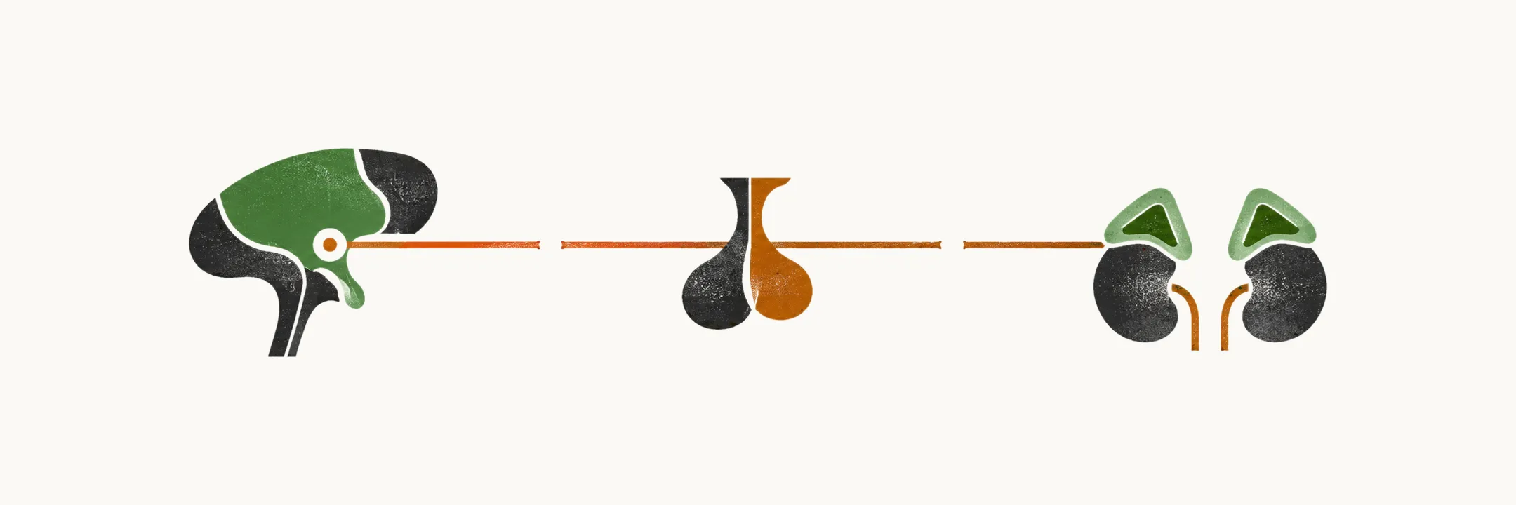
The adrenal cortex is the last link of a chain. The hypothalamus releases CRH, the pituitary answers with ACTH, and ACTH tells the cortex to make cortisol. A chain can fail at any link, so the causes of adrenal insufficiency divide by the level at which the hormone axis fails, and every cause is something that destroys or silences one link.
- Primary form: the adrenal cortex fails.
- Secondary form: the pituitary fails.
- Tertiary form: the hypothalamus fails.
The level matters because it decides which hormones are lost, and that in turn decides what the patient feels and shows.
Who gets primary adrenal insufficiency?
The level here is the gland itself. Primary adrenal insufficiency mostly occurs between ages 30 and 50, and it is more common in women. Its prevalence is 90-140 per million in Europe, and its incidence is 4-6 new cases per million per year. The main cause of primary adrenal insufficiency has changed over time. From the 1950s onward, about 80-90% of cases are due to autoimmune adrenalitis, which is the major cause of cortex destruction in countries where tuberculosis is uncommon.
Why does the immune system attack the cortex?
Autoimmune adrenalitis has been the main cause since the 1950s. The cortex is one of several endocrine glands that the immune system can attack. Autoimmune adrenalitis is isolated in 40% of cases. In the other 60% it is part of an autoimmune polyendocrinopathy syndrome (APS), in which the autoimmunity involves several endocrine glands. The same autoimmune process can therefore reach more than one gland, which is why patients with autoimmune adrenalitis often have other autoimmune diseases.
Addison’s disease is the name for autoimmune adrenalitis. In more than 90% of cases, Addison’s disease is associated with cell-mediated destruction of the adrenal cortex. 21-hydroxylase is an enzyme inside the cortex cells, and the destruction is mediated by autoantibodies against it. Autoantibodies against 21-hydroxylase are reliable markers of this autoimmunity.
Genes shape who is at risk. Genetic polymorphisms predispose to Addison’s disease: HLA-DR3-DQ2 and HLA-DR4-DQ8. Variants of other genes, PTPN22 and CTLA4, also predispose; these are associations, with a smaller effect than the HLA genes, and they do not by themselves cause the disease.
What if an enzyme defect poisons the cortex cells?
An inherited cause damages the same cells without the immune system. Adrenoleukodystrophy is an X-linked disorder that affects the ABCD1 gene. The gene encodes a transporter protein that helps move very long chain fatty acids (VLCFAs) into the peroxisome, and ABCD1 is therefore involved in the β-oxidation of VLCFAs. When this fails, VLCFAs are not broken down and accumulate in the adrenal cortex cells. The accumulation damages the cells and leads to primary adrenal insufficiency; the damage is thought to be cytotoxic. Because the accumulation is mainly in the inner zones of the cortex, mineralocorticoid production is often preserved.
What else destroys the cortex?
Other causes include bleeding or infarction, infection, drugs, tumor spread and infiltration.
Adrenal infarction can occur through hemorrhagic conditions and through adrenal vein thrombosis; both end in loss of cortex tissue. Other causes of hemorrhagic infarction of the adrenals are:
- coagulopathies
- heparin-induced thrombocytopenia
- Waterhouse-Friderichsen syndrome, caused by meningococcemia
- Pseudomonas aeruginosa
Infections reach the adrenal glands and cause adrenalitis and primary adrenal insufficiency. Common infectious causes are:
- Mycobacterium avium complex
- cytomegalovirus infection in HIV-positive patients
- syphilis
Drugs are implicated in drug-induced adrenal insufficiency:
- anti-epileptic drugs, including phenytoin and barbiturates
- sedative drugs
- anti-mycotic drugs
- rifampin
- tyrosine kinase inhibitors
Bilateral adrenal metastasis is another cause of primary adrenal insufficiency, primarily from lung, stomach, breast and colon cancer. It has to be bilateral because most of the cortex has to be lost before insufficiency appears.
Infiltrative disorders are also causes of primary adrenal insufficiency:
- primary bilateral lymphoma of the adrenal gland
- amyloidosis
- hemochromatosis
What goes wrong when the pituitary fails?
Moving up the chain, the lesion in secondary adrenal insufficiency lies in the pituitary and ACTH secretion falls. Secondary adrenal insufficiency is more common than the primary form. Its prevalence is 150-280 cases per million of population, and it mostly occurs in the sixth decade of life in women.
The secondary causes fall into disease categories, each with its mechanism; genetic disorders are named by the mutated gene.
| Category | Cause | Mechanism |
|---|---|---|
| Space-occupying lesions or trauma | Pituitary tumors: adenomas, cysts, craniopharyngiomas, ependymomas, meningiomas, rarely carcinomas | Decreased ACTH secretion |
| Space-occupying lesions or trauma | Trauma (pituitary stalk lesions) | Decreased ACTH secretion |
| Surgery or irradiation | Pituitary surgery or irradiation for pituitary tumors, tumors outside the HPA axis, or leukemia | Decreased ACTH secretion |
| Infections or infiltrative processes | Lymphocytic hypophysitis, hemochromatosis, tuberculosis, meningitis, sarcoidosis, actinomycosis, histiocytosis X, Wegener’s granulomatosis | Decreased ACTH secretion |
| Vascular | Pituitary apoplexy | Decreased ACTH secretion |
| Vascular | Sheehan’s syndrome (peripartum pituitary apoplexy and necrosis) | Decreased ACTH secretion |
A stalk lesion works by the chain logic: a break between the hypothalamus and the pituitary stops the signal from reaching the pituitary.
The genetic causes are named by the mutated gene:
| Disorder | Mechanism |
|---|---|
| HESX homeobox 1 (HESX1) | HESX1 gene mutations |
| Orthodenticle homeobox 2 (OTX2) | OTX2 gene mutations |
| LIM homeobox 4 (LHX4) | LHX4 gene mutations |
| PROP paired-like homeobox 1 (PROP1) | PROP1 gene mutations |
| SRY (sex-determining region Y)-box 3 (SOX3) | SOX3 gene mutations |
| T-box 19 (TBX19) | TBX19 gene mutations |
| Congenital proopiomelanocortin (POMC) deficiency | POMC gene mutations |
| Prader-Willi syndrome (PWS) | Deletion or silencing of genes in the imprinting center for PWS |
What goes wrong when the hypothalamus fails?
One level higher, tertiary adrenal insufficiency is based on low production and secretion of CRH from the hypothalamus. The most common cause is chronic use of synthetic glucocorticoids. The body reads a synthetic glucocorticoid as cortisol, so negative feedback switches off the release of CRH and ACTH and the adrenal cortex is no longer stimulated.
The tertiary causes are grouped by disease category. The drug-induced group includes glucocorticoid therapy, Cushing’s syndrome, and drugs that block glucocorticoid action in tissues. Cushing’s syndrome belongs here because the body itself makes too much glucocorticoid, which suppresses the axis in the same way as glucocorticoid tablets.
| Category | Cause | Mechanism |
|---|---|---|
| Space-occupying lesions or trauma | Hypothalamic tumors (craniopharyngiomas or metastasis from lung or breast cancer) | Decreased CRH secretion |
| Surgery or irradiation | Hypothalamic surgery or irradiation for central nervous system or nasopharyngeal tumors | Decreased CRH secretion |
| Infections or infiltrative processes | Lymphocytic hypophysitis, hemochromatosis, tuberculosis, meningitis, sarcoidosis, actinomycosis, histiocytosis X, Wegener’s granulomatosis | Decreased CRH secretion |
| Trauma | Injury (fracture of the skull base) | Decreased CRH secretion |
| Drug-induced | Glucocorticoid therapy (systemic or topical) or endogenous glucocorticoid hypersecretion (Cushing’s syndrome) | Decreased CRH and ACTH secretion |
| Drug-induced | Mifepristone | Tissue resistance to glucocorticoids through impairment of glucocorticoid signal transduction |
| Drug-induced | Antipsychotics (chlorpromazine), antidepressants (imipramine) | Inhibition of glucocorticoid-induced gene transcription |
The infections and infiltrative processes are the same as those that act on the pituitary, here acting on the hypothalamus. The last two rows reduce the action of glucocorticoids rather than their production, so cortisol can be present while its effect is reduced.
What does the level of the lesion decide?
Whether the lesion sits in the adrenal gland, the pituitary or the hypothalamus decides which hormones are lost, and which hormones are lost decides what the patient feels and shows. Now that the level fixes the hormones lost, what does a patient actually feel and show, and why do the three forms look different at the bedside?