
The adrenal cortex supplies three things the body leans on every minute. Cortisol keeps blood pressure, blood glucose and appetite steady. Aldosterone holds sodium and water in and lets potassium go. Androgens support libido and, in women, much of the drive behind sexual and body-hair features. The pituitary’s ACTH drives cortisol and, through its own precursor, also stimulates pigment cells. When the gland is destroyed, all three hormones are lost and ACTH rises. When the pituitary or hypothalamus fails, cortisol and androgens fall, ACTH falls, and aldosterone is untouched. Everything the patient shows follows from which of these are missing and from how fast they were lost.
What does the slow picture look like, and why is it vague?
Adrenal insufficiency usually develops as a chronic, progressive illness. The clinical picture of the chronic form depends on how much of the adrenal gland is lost and on whether the zona glomerulosa, the outer layer that makes mineralocorticoids, is involved. Partial loss can look well at rest, because the gland first loses its reserve and only later its basal secretion.
Primary adrenal insufficiency is by nature a chronic, progressive condition, and it strips all three hormones: glucocorticoids, mineralocorticoids and androgens. Because so many separate hormone roles are lost together, there is no single telling sign. Instead the patient develops non-specific signs and symptoms that accumulate over time.
The illness can also become acute, as an adrenal crisis. The crisis is chiefly a complication of the primary form, for reasons that become clear once the loss of mineralocorticoid is considered. It can also occur in the secondary and tertiary forms.
What does chronic primary adrenal insufficiency look like?
With all three hormones lost and ACTH high, the picture can be read hormone by hormone.
Glucocorticoid loss. Cortisol deficiency disturbs metabolism and muscle function, which fits the malaise and fatigue of the chronic form. Weight loss is also seen; salt and water loss and low volume are thought to contribute at least in part, and the full mechanism is not fully understood. Nausea and vomiting are common, as is diarrhea alternating with constipation; these gastrointestinal upsets are typical non-specific features of the illness.
Mineralocorticoid loss. Mineralocorticoids stimulate sodium reabsorption and potassium excretion. Without them, sodium and water are lost and potassium is retained, so the patient has hyponatremia and hyperkalemia. Volume loss and dehydration follow, and metabolic acidosis belongs to the same electrolyte imbalance. The loss of sodium, water and circulating volume also produces hypotension, and glucocorticoid deficiency contributes to it as well.
Androgen loss. The cortex normally makes androgens, so their loss produces lack of libido and amenorrhea.
High ACTH and pigment. Low cortisol removes negative feedback, so the pituitary raises ACTH. ACTH comes with beta-lipotropin from the same precursor, and both have melanocyte-stimulating activity, so skin and mucous membranes darken. This is hyperpigmentation of the skin, and it is due to the high ACTH level. Vitiligo can also be present.
The patient who looks like this can suddenly look much worse. How does that happen?
How does a patient with primary adrenal insufficiency slide into crisis?
Primary adrenal insufficiency can be complicated by an acute episode, adrenal crisis. In a crisis, glucocorticoid secretion falls abruptly: the body cannot meet its demand for cortisol. Its major manifestation is shock, because cortisol deficiency contributes to hypotension and reduced cardiac output, while mineralocorticoid deficiency empties the circulating volume. Patients may also have non-specific features: nausea, anorexia, abdominal pain, confusion, weakness and fatigue.
The major precipitating factor of crisis in primary adrenal insufficiency is mineralocorticoid deficiency. Without mineralocorticoids, sodium and water are lost in the urine, causing severe dehydration, lower circulating volume, hypotension and circulatory collapse, and this sodium and water loss dominates the crisis. This explains why crisis mainly happens in patients who are not receiving the mineralocorticoid replacement they need.
A second mechanism is also thought to operate: a low glucocorticoid level desensitizes adrenergic receptors. If so, the patient responds less to adrenergic stimulation, and the resulting fall in vasoconstriction can add to the shock. This is a proposed explanation.
Crisis may occur in response to a major stressor, because the patient has little reserve and cannot raise the extra cortisol the body then needs. The stressors are:
- infections, which raise the demand for cortisol;
- hemorrhage or infarction of the adrenal glands, which removes the remaining cortex abruptly;
- sudden withdrawal of steroid replacement therapy, which removes the only source of cortisol.
The causes of gland destruction, including adrenal hemorrhage, are covered in Causes of Adrenal Insufficiency. If the lesion lies higher up, in the pituitary or hypothalamus, the picture changes.
How do the secondary and tertiary forms look different?
Cortisol and androgens fall in the secondary and tertiary forms as they do in the primary form, so the signs and symptoms are very similar to primary, with two exceptions.
- There is no hyperpigmentation, because ACTH or CRH is deficient rather than high, so there is no excess ACTH or beta-lipotropin to stimulate pigment cells.
- There is no hyperkalemia, metabolic acidosis or dehydration from mineralocorticoid loss, because aldosterone secretion is preserved, unlike in primary adrenal insufficiency.
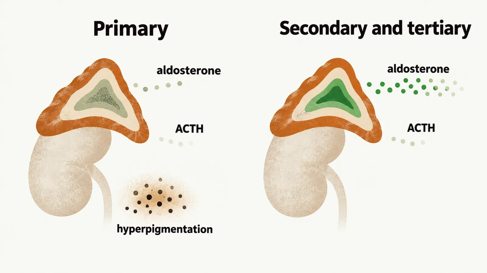
Hyponatremia can nevertheless occur. It happens because cortisol deficiency itself impairs the kidney’s ability to excrete free water, so sodium can fall even when aldosterone is normal. This is a different mechanism from the sodium loss of the primary form.
Depending on the cause and the extent of progression, neurological and hormonal signs and symptoms also appear in the secondary and tertiary forms. They arise from mass effect or from involvement of other anterior pituitary hormones. Headache, hormonal insufficiency and visual disturbances can be seen.
Hypoglycemia is more common in secondary adrenal insufficiency. Cortisol normally supports glucose production, so lack of it lowers glucose. Concomitant lack of growth hormone (GH), which can be seen in secondary adrenal insufficiency, is a possible additional reason.
Does a crisis still happen then?
Adrenal crisis differs in the secondary and tertiary forms. The zona glomerulosa is preserved, so aldosterone secretion continues. These forms are therefore not complicated by the salt and water loss of primary adrenal insufficiency, and a hypovolemic crisis is much less likely.
A crisis can still occur when a major stressor such as infection arrives. The pituitary or hypothalamus cannot raise ACTH, so the cortisol surge the body needs does not come.
Once a doctor suspects any of these pictures, how is the low cortisol confirmed, and how do the hormone patterns show whether the gland, the pituitary or the hypothalamus is at fault?