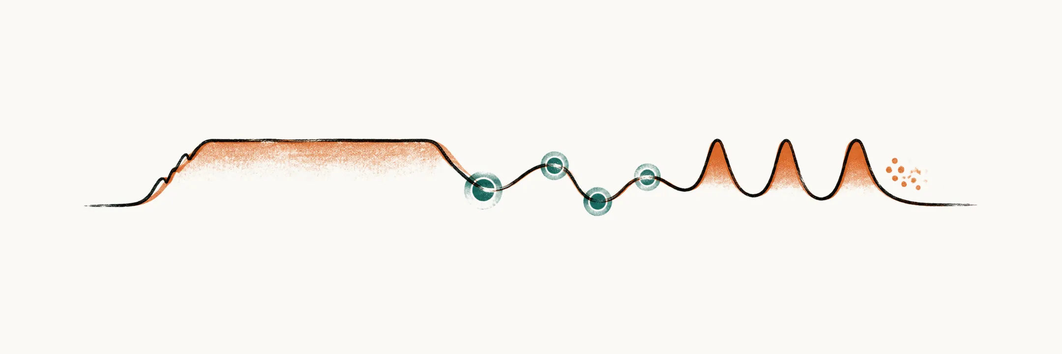
Why do three different diseases look like one?
Kawasaki disease, acute rheumatic fever and juvenile idiopathic arthritis (JIA) can each present as a febrile child with a rash, joint pain or a cardiac finding. The three are frequently confused. Yet each attacks a different tissue.
- Kawasaki disease is a vasculitis of medium-sized arteries in young children, and the coronary arteries are its target.
- Acute rheumatic fever is an immune cross-reaction: the response to a group A streptococcal throat infection also attacks the child’s own tissues.
- JIA is a chronic synovitis. Systemic JIA is instead autoinflammatory, driven by the innate, non-specific arm of the immune system.
Signs such as fever, rash, a swollen joint or a heart finding are shown by many illnesses, so one sign cannot name the disease. What separates the three is where the inflammation sits, how it behaves over days and weeks, and above all what happens to the heart. The decisions that matter are also time-limited. Kawasaki disease needs IVIG within 10 days of fever onset, and a child treated late may carry a coronary aneurysm for life. Rheumatic fever prophylaxis depends on a streptococcal infection being recognised. So what tells them apart?
What separates them?
Each disease attacks a different tissue, so each answers the same few questions differently. The table sets the features side by side.
| Kawasaki disease | Acute rheumatic fever | JIA | |
|---|---|---|---|
| Mechanism | Medium-vessel vasculitis | Immune-mediated cross-reaction after streptococcal pharyngitis | Chronic immune-mediated synovitis; systemic JIA is autoinflammatory |
| Trigger | Unknown, probably infectious in a susceptible child | Rheumatogenic group A Streptococcus | Unknown |
| Typical age | 9-11 months; about 80% are under 5 years | 5-15 years | Oligoarthritis 1-2 years; polyarthritis school age or adolescence; systemic JIA any age |
| Sex | Slight male predominance | Equal; chorea more common in girls | Female predominance in oligoarticular and RF-positive polyarticular disease |
| Fever | Sustained, high, resistant to antipyretics and antibiotics, at least 5 days | A minor criterion; not the presenting focus | Systemic JIA: daily quotidian spikes; other subtypes usually afebrile |
| Rash | Polymorphous maculopapular exanthem | Erythema marginatum (uncommon, under 10%) | Systemic JIA: evanescent pink rash appearing with the fever spike |
| Joints | Sometimes involved, especially in atypical presentations | Migratory, transient, asymmetric, large joints | Persistent; oligoarthritis affects up to 4 joints, polyarthritis at least 5; symmetric small joints when RF-positive |
| Heart | Coronary artery dilation and aneurysm | Valvulitis, mitral valve first; pancarditis; subclinical carditis on Doppler | Pericarditis in severe systemic JIA is the exception |
| Time-critical step | IVIG within 10 days of fever onset | Treat streptococcal pharyngitis within 9 days to prevent a first attack; long-term prophylaxis after an attack | Early, effective disease-modifying treatment before erosive damage |
| Specific prevention | None | Benzathine penicillin G secondary prophylaxis | None |
Which feature do you check first?
The table shows that fever differs most in its shape, so the practical question is which feature is cheapest and first to check. Fever pattern is the most useful bedside discriminator among the features you can check, and it costs nothing to establish. Parents often give a better account of the fever rhythm than a chart does.
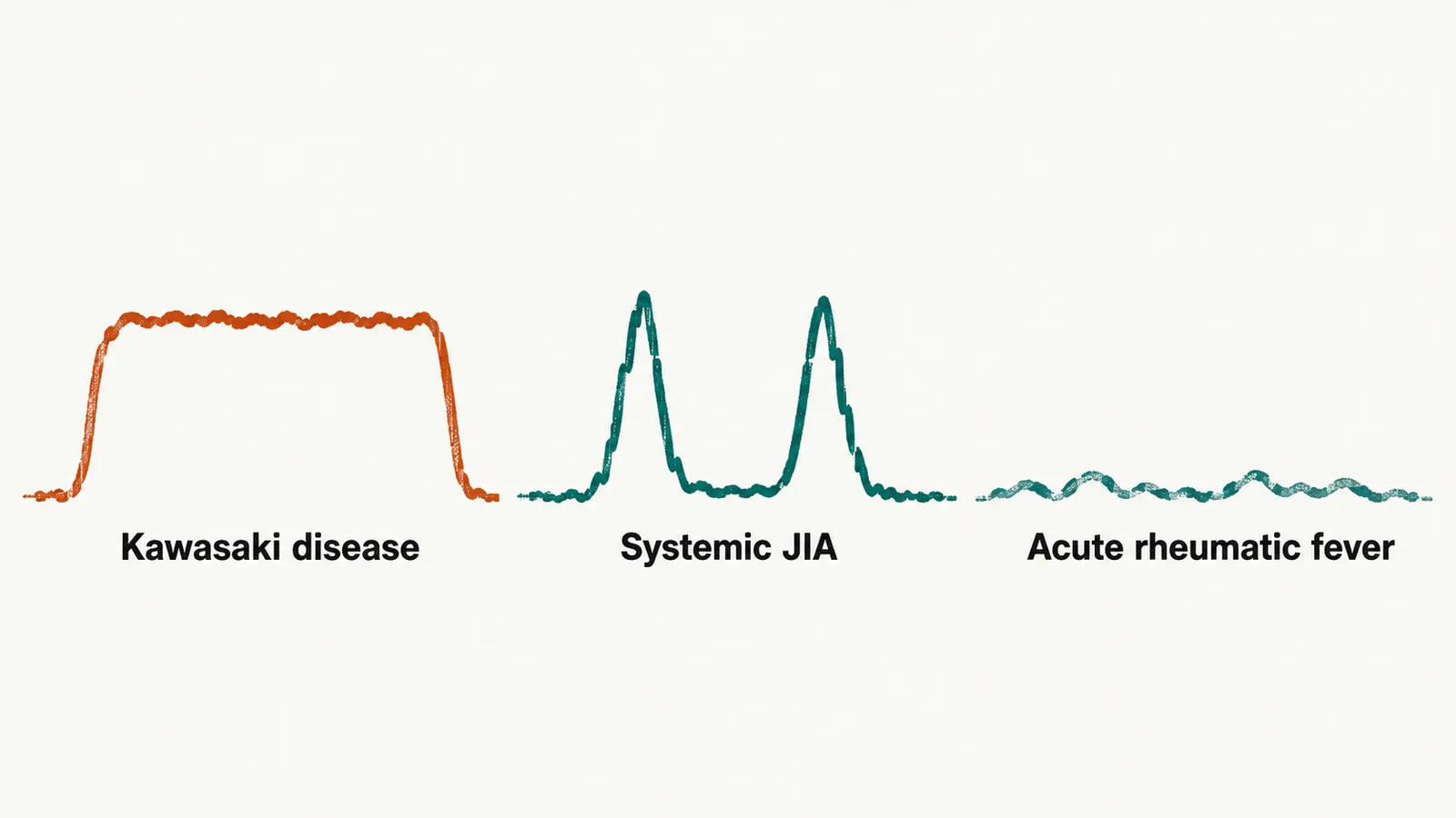
In Kawasaki disease the fever does not respond to antipyretics or antibiotics. It lasts 5 days or more without treatment, and it is continuous rather than spiking.
In systemic JIA there are one or two high spikes every day, and the child returns towards normal in between. This is the quotidian pattern, and the rash appears at the height of each spike.
In acute rheumatic fever, fever is present in most cases, but it is a minor criterion. That means it is a supporting rather than a defining finding in the diagnostic rules. The illness is usually dominated by joint pain.
Persistent fever with no clear focus is also the presentation of serious bacterial infection, urinary tract infection and leukaemia. So the fever pattern is a starting point rather than a conclusion. The fever gives the shape of the illness. What does the rash add?
What does the rash show?
A rash occurs in all three diseases. It is the sign they share, so it is read next and always with its timing, because each rash looks different and appears at a different moment.
The Kawasaki rash is pink and maculopapular, on the trunk and limbs. On its own it is indistinguishable from a viral exanthem. It counts towards the diagnostic criteria, but it is not what identifies the disease.
The rash of acute rheumatic fever is erythema marginatum: non-pruritic reddish rings with a pale centre, on the trunk and proximal limbs, in under 10% of cases. It can be mistaken for the migratory erythema of Lyme disease, and what separates them is exposure history. The ring of Lyme disease is a rash at the site of a tick bite, so a tick history is what points to it. The ring of rheumatic fever follows a sore throat, and there is no bite to ask about.
The rash of systemic JIA is evanescent and salmon-pink, and it comes and goes with the fever spikes. It may be visible only when the temperature is high, so look for it during a spike rather than between spikes.
Why is a red tongue not enough?
A rash that looks like scarlet fever raises the same doubt about the tongue. Strawberry tongue is shared by several illnesses. It occurs in Kawasaki disease, scarlet fever and toxic shock syndrome, so it is not a discriminator by itself. That is why scarlet fever is a differential for all three diseases.
What tells them apart is what accompanies the tongue.
- In Kawasaki disease, other principal features or a coronary abnormality are present. No infecting organism has been identified behind its tongue.
- In scarlet fever there is pharyngitis, a fine sandpaper-like rash accentuated in the skin creases (Pastia’s lines) with circumoral pallor, and a positive streptococcal test.
- In toxic shock syndrome there are shock and multi-organ dysfunction.
What do the joints show over time?
Skin and tongue sort the rash. The joints sort the diagnosis by how they behave over time. Rheumatic fever moves from joint to joint, and the joints recover. In JIA the same joints stay inflamed, and persistent inflammation erodes cartilage and bone. In Kawasaki disease the joints are sometimes involved, especially in atypical presentations.
Some patterns point to a disease.
- Migratory, transient, asymmetric involvement of large joints such as the knee and elbow, after a sore throat, suggests acute rheumatic fever. Migration from one large joint to another over days belongs to rheumatic fever.
- Persistent involvement of up to 4 joints, most often the knee, in a toddler, with ANA positivity suggests oligoarticular JIA.
- Persistent symmetric small-joint disease of the hands and feet in an adolescent girl, with morning stiffness, suggests RF-positive polyarticular JIA. Morning stiffness and symmetric small-joint involvement belong to JIA.
- Arthritis of 5 or more joints within the first 6 months suggests polyarticular JIA.
Chronic, relatively painless monoarticular swelling in a child fits oligoarticular JIA. Tuberculous arthritis and other chronic infection must also be considered.
Two alternatives must not be missed for any joint pattern. A single hot, exquisitely painful joint suggests septic arthritis, and aspiration is required whatever else is considered. Joint pain with bone pain and cytopenia, worst at night, suggests leukaemia, and bone marrow examination is needed.
Joint behaviour separates rheumatic fever from JIA. But Kawasaki disease and rheumatic fever can both damage the heart. What separates them?
What does the heart show?
The two diseases damage different parts of the heart, so the imaging finding tells which disease it is. Kawasaki disease reaches the heart through the coronary artery wall. Rheumatic fever reaches it through the valves and heart muscle.
Kawasaki disease causes coronary dilation or aneurysm, graded by Z-score. A normal echocardiogram early in the illness does not exclude the diagnosis, because aneurysms develop over the following weeks. The echocardiogram is repeated at 1-2 weeks and at 4-6 weeks.
Rheumatic fever causes valvulitis: mitral regurgitation first, then aortic regurgitation. Doppler echocardiography can detect subclinical regurgitation even when no murmur is audible. Pericarditis and myocarditis occur as part of pancarditis, which is inflammation of all three layers of the heart. First-degree atrioventricular block is the common ECG finding.
Systemic JIA causes pericarditis in its severe form, and rarely myocarditis. Coronary aneurysms are not part of its picture.
Echocardiography is therefore the single most useful discriminator in a febrile child with possible cardiac involvement. It is indicated both when Kawasaki disease is suspected and when rheumatic fever is suspected. Can a blood test do the same?
What do the blood tests add?
No single blood test separates the three diseases. Tests are most useful when read with the clinical pattern. CRP and ESR are general markers of inflammation. They are raised in Kawasaki disease and in active rheumatic fever, and both may be normal in oligoarticular JIA.
Some tests point to one disease.
- A rising ASO or anti-DNase B titre on serial samples indicates a preceding group A streptococcal infection. A single raised value is common in healthy school-age children. A normal value does not exclude rheumatic fever, because titres are raised in roughly 85% of patients.
- A positive throat culture or rapid antigen test indicates streptococcal pharyngitis, but these are often negative by the time rheumatic fever appears.
- Platelets rising after day 7 of fever support Kawasaki disease retrospectively.
- Sterile pyuria suggests Kawasaki disease, and it is useful for separating it from a urinary tract infection.
- ANA is positive in roughly three-quarters of oligoarticular JIA, and it marks a higher risk of chronic anterior uveitis.
- Rheumatoid factor defines the RF-positive polyarticular category only when it is positive on two occasions at least 3 months apart.
Blood tests cannot separate the three, but two combinations point to alternatives that must not be missed. Pancytopenia with a very high ferritin, high triglycerides and low fibrinogen suggests macrophage activation syndrome until proven otherwise. Pancytopenia with blasts and bone pain suggests leukaemia until proven otherwise.
Which children do not fit the comparison?
Some children fall outside the comparison altogether, and each needs a different first step. Three situations stand out, and the first priority differs in each.
The first is the infant under 6 months with prolonged fever. This is the group in which Kawasaki disease is most often missed, because infants present with fewer features. An infant of 6 months or younger with fever for 7 days or more and no other explanation should have laboratory testing. If there is evidence of systemic inflammation, the infant should have an echocardiogram even with no principal features. Urinary tract infection and serious bacterial infection must be excluded in parallel.
The second is the child with a single inflamed joint: aspirate first. Septic arthritis, osteomyelitis and, less acutely, tuberculous arthritis all present this way, and a single inflamed joint belongs to septic arthritis until proven otherwise. A joint infection can destroy cartilage within days. A chronic, monoarticular, relatively indolent presentation makes oligoarticular JIA likely. But JIA is a diagnosis of exclusion, so the exclusion list still has to be worked through.
The third is multisystem inflammatory syndrome in children, a distinct post-infectious illness following SARS-CoV-2 infection. Its features are persistent fever, gastrointestinal symptoms, rash, conjunctivitis, myocardial dysfunction and, in some children, coronary involvement. It overlaps substantially with Kawasaki disease, and it is managed differently. It is distinguished by age, by the epidemiological link to a preceding infection, and by the pattern of organ involvement, with more frequent gastrointestinal and myocardial dysfunction and more marked inflammation. So a child meeting Kawasaki criteria today may not have the classic disease.
In what order do you work?
- Document the fever pattern and duration, and examine the whole child: eyes, mouth, hands and feet, neck, skin, joints and heart.
- Look for streptococcal infection with a throat swab or rapid test. Send serology (ASO and anti-DNase B) with a plan to repeat it in 2-3 weeks.
- Obtain an echocardiogram if Kawasaki disease or rheumatic fever is possible. It is the test that separates the two.
- Send inflammatory and supportive tests: full blood count, CRP and ESR, liver enzymes, albumin and urinalysis. They serve the Kawasaki algorithm and give a baseline for the others.
- Do not wait for a titre before treating Kawasaki disease if the clinical criteria are met. A titre takes weeks to rise, while IVIG is time-critical within 10 days of fever onset, so the serology result will not change the decision.
- Consider bone marrow examination and a blood film if fever, cytopenia or bone pain suggest leukaemia.
Consider macrophage activation syndrome if the fever becomes continuous and the counts fall. Once the steps are done, which of the three is it, and how much time does the child have before that disease’s deadline runs out?