
What kind of arthritis is this?
Juvenile idiopathic arthritis (JIA) is a group of categories, and the category decides which threat matters most. In systemic JIA the disease is truly systemic rather than primarily articular. The joints are only part of the illness.
Why is the whole child ill? Systemic JIA is increasingly regarded as autoinflammatory rather than a classic autoimmune disease. It is driven by the innate immune system, the body’s first-line, non-specific defence, and by its cytokines, including IL-1 and IL-6. The child has fever and rash and looks unwell, not just sore joints.
This also explains a treatment fact. Because the drive runs through IL-1 and IL-6, blocking those two cytokines works particularly well in this subtype.
A drive that acts through the whole body explains the next question: why does the fever look like an infection?
Why does the fever look like infection?
Systemic JIA is the JIA category that most often resembles an infection. Fever is the key discriminator.
The fever is typically quotidian. It spikes once or twice daily to high values and returns towards normal in between. The rash follows the same rhythm. It is an evanescent rash: pale pink and non-fixed, it appears with the fever spike and fades as the temperature falls.
Its signal comes from the body’s own innate drive, not from a germ.
The fever often precedes the arthritis by weeks. A child with recurring daily spikes, a rash appearing with each spike and joint pain is a different diagnostic problem from a child with a sustained fever. The shape of the fever chart is often the first clue that the illness is not an ordinary infection.
A fever that looks like infection still has to be told apart from other causes.
How is it told apart from other causes?
Like every JIA category, systemic JIA is diagnosed by recognising a pattern and excluding alternatives. No single test makes the diagnosis. The pattern is arthritis plus fever for at least 2 weeks, plus at least one of the following:
- an evanescent rash
- hepatomegaly or splenomegaly
- generalised lymphadenopathy
- serositis, which is inflammation of the lining of the heart, lungs or abdomen and is most often pericarditis
Because the fever often comes before the arthritis, the alternatives are many. Persistent fever with rash and joint complaints in a child has a wide and serious differential:
- Infection: sepsis, endocarditis, Epstein-Barr virus, cytomegalovirus, osteomyelitis.
- Kawasaki disease: it also gives fever, rash and lymphadenopathy. But its fever is sustained rather than spiking, and the echocardiogram shows coronary involvement.
- Other rheumatic disease: systemic lupus erythematosus and other vasculitides.
- Inflammatory bowel disease with associated arthritis.
The trap in this differential is malignancy. Leukaemia and lymphoma can produce the same picture, especially with bone pain, night pain or cytopenia (low count of one or more blood cell lines). Acute lymphoblastic leukaemia is the classic mimic. Bone marrow examination is part of the workup of a child whose systemic JIA does not behave typically.
Once the pattern is clear, the risk to think about is what the disease can turn into.
Why can it become an emergency?
Macrophage activation syndrome (MAS) is the most dangerous complication of systemic JIA, and it can become fatal within days. It is uncontrolled activation of T lymphocytes and macrophages. Its features overlap with haemophagocytic lymphohistiocytosis (HLH).
MAS can develop at disease onset or during a flare. It is often triggered by an infection or by a change in treatment.
The picture is a child who suddenly becomes much worse:
- unremitting, sustained fever, losing the quotidian pattern
- pancytopenia, a fall in red cells, white cells and platelets together
- hepatosplenomegaly, sometimes with liver failure
- coagulopathy with bleeding
- neurological symptoms: drowsiness, irritability, seizures
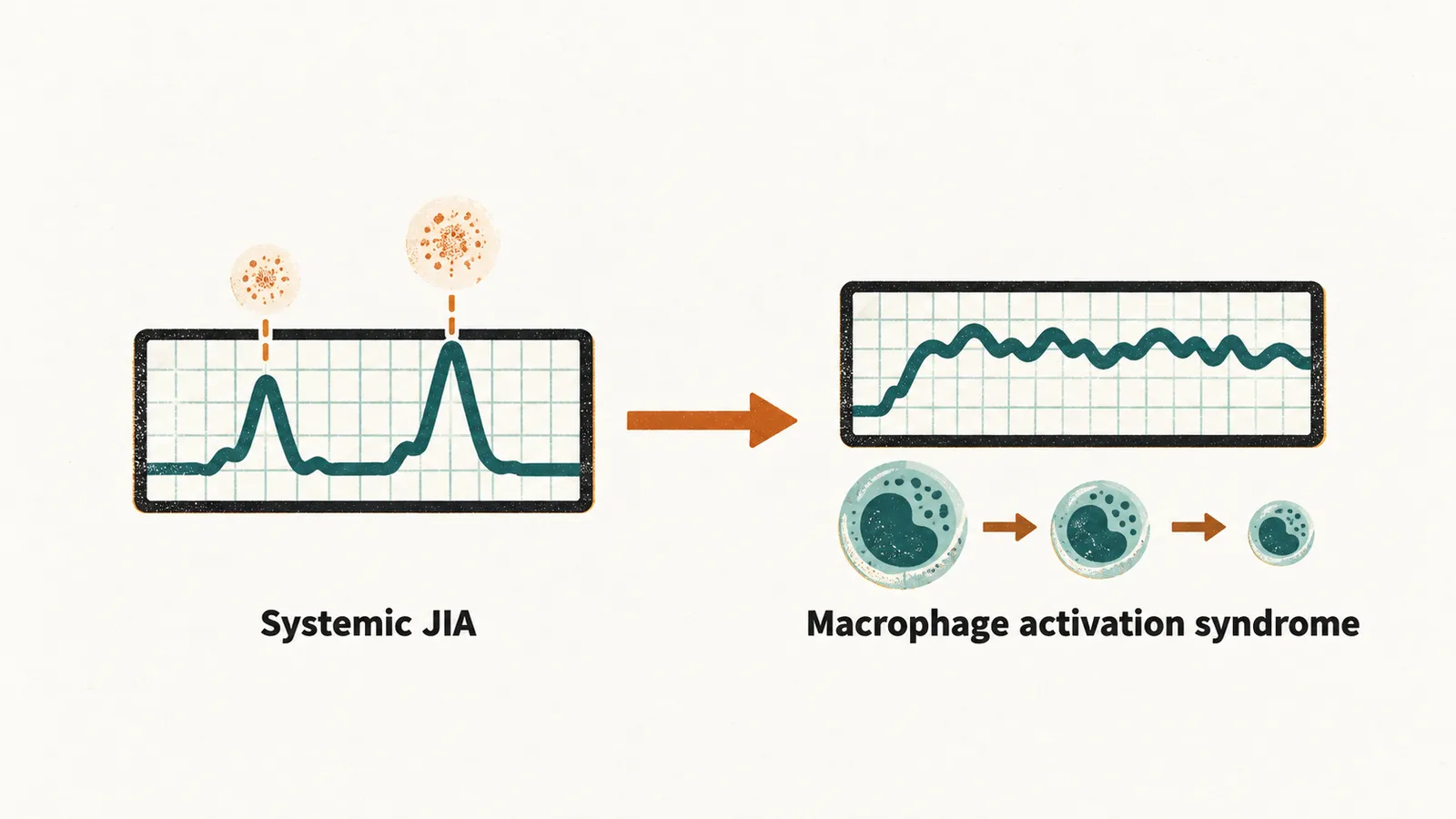
The emergency shows up in the blood, so the next question is what the blood shows.
What do the blood tests show?
The laboratory picture of MAS is:
- very high ferritin
- raised triglycerides
- low fibrinogen
- raised liver enzymes and lactate dehydrogenase
- hyponatraemia
- haemophagocytosis, macrophages that have engulfed blood cells, on bone marrow examination
CRP and ESR are general markers of inflammation, but they do not move together here. A falling ESR in the face of a rising CRP is a useful clue in MAS. The ESR falls because fibrinogen is consumed.
The blood picture shows the storm, and the storm is treated next.
How is MAS treated, and what sets it off?
MAS is a medical emergency with high mortality if it is not recognised. The trigger to remember is a child with known systemic JIA whose fever suddenly becomes continuous and whose blood counts fall.
MAS is treated with high-dose intravenous corticosteroids together with cyclosporine. Both act on T lymphocytes, which are one of the two cell types driving the storm. Cyclosporine suppresses T cell activation, and it may inhibit the production of IL-2 and the expression of its receptor on T lymphocytes.
In refractory cases anakinra, an IL-1 inhibitor, is added.
A treatment that works now still has to be kept up, so the last question is how the disease is held quiet.
How is the disease kept quiet between emergencies?
Between emergencies, systemic disease and the other JIA categories are managed by a long-term strategy measured against a target, treat-to-target, rather than by the severity of the moment. How is that target set, and what drugs reach it?